Lightning-Link® Alexa Fluor® 647 抗體標定試劑盒

Alexa Fluor® 647 Conjugation Kit (Fast) - Lightning-Link®

貨號

ab269823

包裝

3 x 10 μg
100 μg
3 x 100 μg
1 mg

廠牌

Abcam

產品簡介

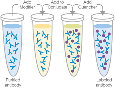
Abcam Alexa Fluor® 647 Conjugation Kit (Fast) - Lightning-Link® 提供了一種簡便快速的方法,可將 Alexa Fluor® 647 螢光染料標定至抗體或其他含有游離胺基的蛋白質、胜肽分子上,標定產物可直接應用於 Western Blot、流式細胞分析 (flow cytometry)、ELISA、免疫組織化學染色 (IHC) 等實驗。套組採用 Lightning-Link® 直接標定技術,僅需將修飾劑 (Modifier) 加入待標定抗體,再以此溶液回溶凍乾的 Alexa Fluor® 647 試劑小瓶,即可啟動標定反應;動手操作時間約 30 秒,20 分鐘左右即可完成標定,全程無需任何純化步驟,抗體回收率可達 100%。
Abcam Lightning-Link® Conjugation Kit

主要特色

  • 操作極為快速,動手操作時間僅約 30 秒,全程約 20 分鐘即可完成。
  • 全程無需純化步驟,抗體回收率可達 100%,標定完成後即可直接應用於 Western Blot、Flow cytometry、ELISA、IHC 等實驗,無需額外處理。
Immunocytochemistry. Direct immunofluorescent staining using Recombinant Anti-Cytokeratin 19 antibody [EP1580Y] - BSA and Azide free (ab195872) labelled with Alexa Fluor® 647 Conjugation Kit (Fast) - Lightning-Link® (ab269823). Lightning-Link® Alexa Fluor® 647 Anti-Cytokeratin 19 [EP1580Y] conjugate was used to stain wild-type MCF7 cells (top panel) and A431 negative cells (bottom panel). The cells were fixed with 4% paraformaldehyde (10 min) or 100% methanol at -20°C (5min), permeabilized with 0.1% Triton X-100 for 5 minutes and blocked with 1% BSA/10% normal goat serum/0.3M glycine in 0.1% PBS-Tween for 1h. The cells were incubated with 1 μg/ml of LL-Anti-Cytokeratin 19 Alexa Fluor® 647 conjugate (shown in red) or ab195872 (Anti-Cytokeratin 19 antibody, unlabelled control) overnight at +4°C. ab195872 treated cells only were incubated with ab150077 at 1/1000 dilution for 1 hour at room temperature (shown in green). Nuclear DNA was labelled in blue with DAPI. Image was taken with a confocal microscope (Leica-Microsystems, TCS SP8).
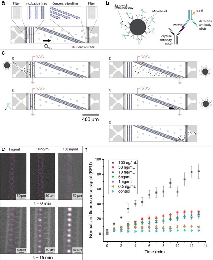
Immunoelectrophoresis. Thiriet, Pierre-Emmanuel, et al used Alexa Fluor® 647 Conjugation Kit (Fast) - Lightning-Link® (ab269823) as part of rapid and accurate diagnosis of Acute kidney injury (AKI). They used the kit to conjugate Alexa Fluor® 647 to monoclonal anti-Lipocalin-2/NGAL antibody for use in the development of a microfluidic analytical device . (a-c) Chip description and operation. (a) Presentation of the chip layout. Beads and reagents can be successively injected through the two inlets visible on the left. The device consists of three incubation lines upstream and three concentration lines downstream, at the end of which the beads are accumulated in clusters (shown here in red). (b) Illustration of a sandwich immunoassay used for detection of biomarkers. The analyte we aimed to detect was captured by the bead decorated with capture antibody (cAb) and detection was performed thanks to the fluorescently labeled detection antibody (dAb). (c) Presentation of the successive steps performed on-chip to operate the platform, namely, (1) beads' loading, (2) incubation with detection antibodies and (3) release from the incubation line, (4) clustering in the concentration region, and (5) discarding through the outlet. For the sake of clarity, the species bound to the beads and the electrically activated arrays of electrodes are indicated for each step. (d-f) On-chip incubation of Neutrophil Gelatinase-Associated Lipocalin (NGAL) biomarker. (d) Observation of the small beads' clusters (circled in pink) before and after 15 min of incubation. The fluorescence signal arose from the binding of dAb-NGAL complex to cAb-decorated beads dielectrically trapped in the regions upstream to the electrode line. Three NGAL concentrations were injected in separate experiments, namely, 1 ng/mL, 10 ng/mL, and 100 ng/mL. (f) Fluorescence signal as a function of the incubation time for different NGAL concentrations. After 15 min all concentrations provided a signal greater than the control experiment, consisting of an injection of a solution in absence of NGAL molecules. The error bars were obtained by measuring the fluorescent signal from 10 clusters.
Immunoelectrophoresis. Thiriet, Pierre-Emmanuel, et al used Alexa Fluor® 647 Conjugation Kit (Fast) - Lightning-Link® (ab269823) as part of rapid and accurate diagnosis of Acute kidney injury (AKI). They used the kit to conjugate Alexa Fluor® 647 to monoclonal anti-Lipocalin-2/NGAL antibody for use in the development of a microfluidic analytical device . (a-c) Chip description and operation. (a) Presentation of the chip layout. Beads and reagents can be successively injected through the two inlets visible on the left. The device consists of three incubation lines upstream and three concentration lines downstream, at the end of which the beads are accumulated in clusters (shown here in red). (b) Illustration of a sandwich immunoassay used for detection of biomarkers. The analyte we aimed to detect was captured by the bead decorated with capture antibody (cAb) and detection was performed thanks to the fluorescently labeled detection antibody (dAb). (c) Presentation of the successive steps performed on-chip to operate the platform, namely, (1) beads' loading, (2) incubation with detection antibodies and (3) release from the incubation line, (4) clustering in the concentration region, and (5) discarding through the outlet. For the sake of clarity, the species bound to the beads and the electrically activated arrays of electrodes are indicated for each step. (d-f) On-chip incubation of Neutrophil Gelatinase-Associated Lipocalin (NGAL) biomarker. (d) Observation of the small beads' clusters (circled in pink) before and after 15 min of incubation. The fluorescence signal arose from the binding of dAb-NGAL complex to cAb-decorated beads dielectrically trapped in the regions upstream to the electrode line. Three NGAL concentrations were injected in separate experiments, namely, 1 ng/mL, 10 ng/mL, and 100 ng/mL. (f) Fluorescence signal as a function of the incubation time for different NGAL concentrations. After 15 min all concentrations provided a signal greater than the control experiment, consisting of an injection of a solution in absence of NGAL molecules. The error bars were obtained by measuring the fluorescent signal from 10 clusters. Image from Thiriet, Pierre-Emmanuel, et al., Biosensors, 10(12):212; doi: 10.3390/bios10120212. Reproduced under the Creative Commons license https://creativecommons.org/licenses/by/4.0/
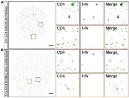
Single-molecule super-resolution imaging. Yuan, Yue, et al used Alexa Fluor 647 Conjugation Kit (Fast) - Lightning-Link (ab269823) as part of examining changes in the nanoscale organisation of CD4 on the surface of CD4+ T cells following HIV-1 binding. They used the kit to conjugate Alexa Fluor 647 to anti-CD4 antibody (OKT4) for use in single-molecule super-resolution imaging. Representative TIRF-STORM images, and selected magnified regions (insets) of cell-surface CD4 (green) and HIV p24 (magenta). Scale bar = 2 µm.
Single-molecule super-resolution imaging. Yuan, Yue, et al used Alexa Fluor 647 Conjugation Kit (Fast) - Lightning-Link (ab269823) as part of examining changes in the nanoscale organisation of CD4 on the surface of CD4+ T cells following HIV-1 binding. They used the kit to conjugate Alexa Fluor 647 to anti-CD4 antibody (OKT4) for use in single-molecule super-resolution imaging. Representative TIRF-STORM images, and selected magnified regions (insets) of cell-surface CD4 (green) and HIV p24 (magenta). Scale bar = 2 µm. Image from Yuan et al., Viruses, 13(1):142. doi: 10.3390/v13010142. Reproduced under the Creative Commons license https://creativecommons.org/licenses

實驗注意事項

  • 樣本緩衝液 pH 值須介於 6.5–8.5,相容成分包括:Tris(濃度應 ≤50 mM / 0.6%)、BSA(≤0.1%)、甘油 (glycerol,≤50%)、疊氮化鈉 (Sodium Azide,≤0.1%)、磷酸鉀、氯化鈉、蔗糖、檸檬酸鈉、海藻糖等。緩衝液不可含有 Thimerosal (Thiomersal)、Proclin、Glycine、Arginine、Glutathione 等成分,以免干擾標定反應。更多樣本緩衝液細部條件說明,可參照下方表格或產品說明書。
  • 僅適用於已純化的抗體樣本;若樣本中含有其他蛋白質、胜肽或游離胺基分子,可能會競爭消耗標定試劑,影響標定效率。抗體樣本理想濃度為 1 mg/mL,若濃度不足(< 0.5 mg/ml)或過高(> 2 mg/ml),建議先進行濃縮或稀釋處理。
  • 標定完成的產物於 4°C 保存可穩定達 18 個月;若需長期保存,建議加入 50% 甘油作為冷凍保護劑,並存放於 -20°C。實際最適合的保存條件仍視個別抗體的穩定性而定,建議透過實驗確認。
Compatible buffer constituents
50mM / 0.6% Tris10.1% BSA250% glycerol
0.1% sodium azidePBSPotassium phosphate
Sodium chlorideHEPESSucrose
Sodium citrateEDTATrehalose
1 Tris buffered saline is almost always ≤ 50 mM / 0.6%
2 BSA can also interfere with the use of the conjugated antibody in tissue staining.
Incompatible buffer constituents
ThiomerosalProclinGlycine
ArginineGlutathioneDTT

套組內容物

Item3 x 10 µg100 µg3 x 100 µg1 mgStorage temperature
Alexa Fluor® 647 mix3 vials1 vial3 vials1 vial-20°C
Modifier reagent1 vial1 vial1 vial1 vial-20°C
Quencher reagent1 vial1 vial1 vial1 vial-20°C

產品說明書

ab269823 Alexa Fluor® 647 Conjugation Kit (Fast) - Lightning-Link®
返回頂端