Lightning-Link® Biotin 抗體標定試劑盒

Biotinylation Kit / Biotin Conjugation Kit (Fast, Type A) - Lightning-Link®

貨號

ab201795

包裝

3 x 10 μg
100 μg
3 x 100 μg
1 mg

廠牌

Abcam

產品簡介

Abcam Biotin Conjugation Kit (Fast, Type A) - Lightning-Link® 提供了一種簡便快速的方法,可將生物素 (Biotin) 標定至抗體或其他含有游離胺基的蛋白質、胜肽分子上,標定產物可直接應用於 Western Blot、流式細胞分析 (flow cytometry)、ELISA、免疫組織化學染色 (IHC) 等實驗。套組採用 Lightning-Link® 直接標定技術,僅需將修飾劑 (Modifier) 加入待標定抗體,再以此溶液回溶凍乾的 Biotin 試劑小瓶,即可啟動標定反應;動手操作時間約 30 秒,20 分鐘左右即可完成標定,全程無需任何純化步驟,抗體回收率可達 100%。
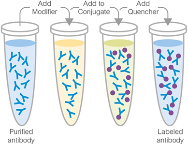
Abcam Lightning-Link® Conjugation Kit

主要特色

  • 操作極為快速,動手操作時間僅約 30 秒,全程約 20 分鐘即可完成。
  • 全程無需純化步驟,抗體回收率可達 100%,標定完成後即可直接應用於 Western Blot、Flow cytometry、ELISA、IHC 等實驗,無需額外處理。
  • 本套組(Type A 版本)適用於以 Streptavidin 標定的偵測試劑進行後續偵測的實驗設計(例如以 Streptavidin-HRP、Streptavidin-螢光染料作為二次偵測試劑);若您的實驗是以固定於平面或磁珠表面的 Streptavidin 捕捉生物素標記蛋白質 (biotinylated protein),如 ELISA 捕捉型偵測、磁珠純化等,建議改選用 Type B 版本 (ab201796)。
ELISA. Direct ELISA dose response curve generated with varying concentration of biotin conjugated antibodies. Anti-MMP1 antibody [EPR19769-152] - BSA and Azide free (Detector) (ab242835), Anti-EGF antibody [EPR19899-46] - BSA and Azide free (Detector) (ab242990),Anti-IL-5 antibody [EPR20012-256] - BSA and Azide free (Capture) (ab242955) and Anti-MMP9 antibody [EPR18543-47] - BSA and Azide free (Capture) (ab242950) were conjugated to biotin using Biotinylation Kit / Biotin Conjugation Kit (Fast, Type A) - Lightning-Link® (ab201795) to produce Conjugate A, B, C and Control respectively. Briefly, each well was coated with Recombinant Protein G. After blocking with 5% BSA blocking buffer for 2 hours, plates were coated with 1ug/ml of biotin conjugated antibodies. Detection was performed using a HRP-conjugated Streptavidin (ab7403), and visualised using a TMB substrate. Plates were read at 450nm using an Agilent BioTek Synergy HTX multimode reader.
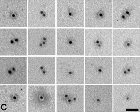
Electron Microscopy. Hansen, Uwe, et al used Biotinylation Kit / Biotin Conjugation Kit (Fast, Type A) - Lightning-Link® (ab201795) as part of examining WARP-collagen IV interactions in in vitro in cartilage. They used the kit to conjugate biotin to recombinant WARP for use in Electron Microscopy. Biotinylated recombinant WARP was visualized by 5 nm gold labeled with streptavidin. Scale bar is 25 nm.
Electron Microscopy. Hansen, Uwe, et al used Biotinylation Kit / Biotin Conjugation Kit (Fast, Type A) - Lightning-Link® (ab201795) as part of examining WARP-collagen IV interactions in in vitro in cartilage. They used the kit to conjugate biotin to recombinant WARP for use in Electron Microscopy. Biotinylated recombinant WARP was visualized by 5 nm gold labeled with streptavidin. Scale bar is 25 nm. Image from Hansen, Uwe, et al., Plos one, 7(12): e52793.; doi: 10.1371/journal.pone.0052793. Reproduced under the Creative Commons license https://creativecommons.org/licenses/by/4.0/
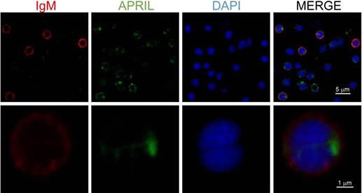
Fluorescence Microscopy. Soleto, Irene, et al used Biotinylation Kit / Biotin Conjugation Kit (Fast, Type A) - Lightning-Link® (ab201795) as part of examining the effect of APRIL on B cells using rainbow trout (Oncorhynchus mykiss) as a model species. They used the kit to conjugate biotin to a recombinant trout proliferation-inducing ligand (APRIL) for use in Fluorescence microscopy. Total leukocytes from spleen were incubated with recombinant biotinylated APRIL (1 μg/ml) for 15 min, then plated onto poly-l-lysine-coated glass slides, fixed and labeled with anti-IgM (shown as red) and FITC-streptavidin (APRIL, shown as green), then counterstained with DAPI (blue), and analyzed by confocal fluorescence microscopy.
Fluorescence Microscopy. Soleto, Irene, et al used Biotinylation Kit / Biotin Conjugation Kit (Fast, Type A) - Lightning-Link® (ab201795) as part of examining the effect of APRIL on B cells using rainbow trout (Oncorhynchus mykiss) as a model species. They used the kit to conjugate biotin to a recombinant trout proliferation-inducing ligand (APRIL) for use in Fluorescence microscopy. Total leukocytes from spleen were incubated with recombinant biotinylated APRIL (1 μg/ml) for 15 min, then plated onto poly-l-lysine-coated glass slides, fixed and labeled with anti-IgM (shown as red) and FITC-streptavidin (APRIL, shown as green), then counterstained with DAPI (blue), and analyzed by confocal fluorescence microscopy. Image from Soleto, Irene, et al., Frontiers in immunology, 9:1880. doi: 10.3389/fimmu.2018.01880; Reproduced under the Creative Commons license https://creativecommons.org/licenses/by/4.0/
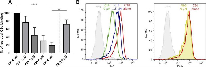
Flow Cytometry. Giussani, Stefania, et al used Biotinylation Kit / Biotin Conjugation Kit (Fast, Type A) - Lightning-Link® (ab201795) as part of examining interaction of C3d ligand with complement interfering protein (CIP). They used the kit to conjugate biotin to C3d for use in flow cytometry. Flow cytometry analysis of C3d interaction with B cells in presence or absence of Complement Interfering Protein (CIP). A) Biotinylated C3d was preincubated with SA and then with 1–9 μM of CIP, 9 μM of Fib3, or buffer alone. Each mixture was added to Raji B cells and treated as described. Binding of the biotinylated C3d–SA complex to cells was revealed by flow cytometry using a C3d-specific mAb and a phycoerythrin-labeled secondary antibody. Mean fluorescence intensity values of the peaks were analyzed by the FlowJo software. The graph shows the percentage of the residual mean fluorescence intensity values in the presence of a competitor compared with buffer alone, as derived from 4 independent experiments. B) Flow cytometry analysis of C3d binding to enriched B cells from human PBMCs in presence or absence of 1.5 or 9 μM CIP or 9 μM of Fib3; experimental conditions were the same as in A.
Flow Cytometry. Giussani, Stefania, et al used Biotinylation Kit / Biotin Conjugation Kit (Fast, Type A) - Lightning-Link® (ab201795) as part of examining interaction of C3d ligand with complement interfering protein (CIP). They used the kit to conjugate biotin to C3d for use in flow cytometry. Flow cytometry analysis of C3d interaction with B cells in presence or absence of Complement Interfering Protein (CIP). A) Biotinylated C3d was preincubated with SA and then with 1–9 μM of CIP, 9 μM of Fib3, or buffer alone. Each mixture was added to Raji B cells and treated as described. Binding of the biotinylated C3d–SA complex to cells was revealed by flow cytometry using a C3d-specific mAb and a phycoerythrin-labeled secondary antibody. Mean fluorescence intensity values of the peaks were analyzed by the FlowJo software. The graph shows the percentage of the residual mean fluorescence intensity values in the presence of a competitor compared with buffer alone, as derived from 4 independent experiments. B) Flow cytometry analysis of C3d binding to enriched B cells from human PBMCs in presence or absence of 1.5 or 9 μM CIP or 9 μM of Fib3; experimental conditions were the same as in A. Image from Giussani, Stefania, et al., The FASEB Journal 33.3 (2019): 4448-4457. Reproduced under the Creative Commons license https://creativecommons.org/licenses/by/4.0/
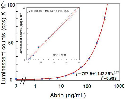
ELISA. He, Xiaohua, et al used Biotinylation Kit / Biotin Conjugation Kit (Fast, Type A) - Lightning-Link® (ab201795) as part of examining the detection of abrin by monoclonal antibodies. They used the kit to conjugate biotin to anti-Abrin antibody for use in ELISA. Detection of abrin in PBS by ELISA. Data represent the average of three determinations ± SD. Limit of detection (LOD) for the mixture of abrin was 1 ng/mL. The one-sided 95% confidence intervals on the fitted line are shown as dashed curves.
ELISA. He, Xiaohua, et al used Biotinylation Kit / Biotin Conjugation Kit (Fast, Type A) - Lightning-Link® (ab201795) as part of examining the detection of abrin by monoclonal antibodies. They used the kit to conjugate biotin to anti-Abrin antibody for use in ELISA. Detection of abrin in PBS by ELISA. Data represent the average of three determinations ± SD. Limit of detection (LOD) for the mixture of abrin was 1 ng/mL. The one-sided 95% confidence intervals on the fitted line are shown as dashed curves. Image from He, Xiaohua, et al., Toxins 9.12 (2017): 386. Reproduced under the Creative Commons license https://creativecommons.org/licenses/by/4.0/
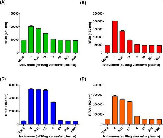
ELISA. Liu, Chien-Chun, et al used Biotinylation Kit / Biotin Conjugation Kit (Fast, Type A) - Lightning-Link® (ab201795) as part of examining assays for snakebite detection based on clinical antivenom usage. They used the kit to conjugate biotin to neurotoxic species-specific antibodies (NSS-Abs) and hemorrhagic species-specific antibodies (HSS-Abs) for use in ELISA. (A, B) Venom proteins of (A) Trimeresurus stejnegeri and (B) Protobothrops mucrosquamatus were diluted in human plasma (10 ng venom protein per ml of plasma) and then mixed with serially diluted Freeze-dried hemorrhagic antivenom (FHAV) (0.32 to 1000 nl) at room temperature for 30 min. The mixtures were then subjected to HSS-Ab–based sandwich ELISA assay. (C, D) Venom proteins of (C) Bungarus multicinctus and (D) Naja atra were diluted in human plasma (10 ng venom protein per ml of plasma) and then mixed with serially diluted FHAV (0.32 to 1000 nl) at room temperature for 30 min. The mixtures were then subjected to NSS-Ab–based sandwich ELISA assay.
ELISA. Liu, Chien-Chun, et al used Biotinylation Kit / Biotin Conjugation Kit (Fast, Type A) - Lightning-Link® (ab201795) as part of examining assays for snakebite detection based on clinical antivenom usage. They used the kit to conjugate biotin to neurotoxic species-specific antibodies (NSS-Abs) and hemorrhagic species-specific antibodies (HSS-Abs) for use in ELISA. (A, B) Venom proteins of (A) Trimeresurus stejnegeri and (B) Protobothrops mucrosquamatus were diluted in human plasma (10 ng venom protein per ml of plasma) and then mixed with serially diluted Freeze-dried hemorrhagic antivenom (FHAV) (0.32 to 1000 nl) at room temperature for 30 min. The mixtures were then subjected to HSS-Ab–based sandwich ELISA assay. (C, D) Venom proteins of (C) Bungarus multicinctus and (D) Naja atra were diluted in human plasma (10 ng venom protein per ml of plasma) and then mixed with serially diluted FHAV (0.32 to 1000 nl) at room temperature for 30 min. The mixtures were then subjected to NSS-Ab–based sandwich ELISA assay. Image from Liu, Chien-Chun, et al., PLoS Negl Trop Dis;12(12): e0007014. doi: 10.1371/journal.pntd.0007014. Reproduced under the Creative Commons license https://creativecommons.org/licenses/by/4.0/
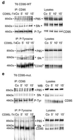
Western Blot. Drachsler, Moritz, et al used Biotinylation Kit / Biotin Conjugation Kit (Fast, Type A) - Lightning-Link® (ab201795) as part of examining expression of CD95. They used the kit to conjugate biotin to anti-human CD95 for use in Western blot. IP for CD95 and P-Tyrosine GSCs in naive, CD95-WT and CD95-mut GBM cells stimulated with CD95L-T4. Blots were probed with anti-P85 (regulatory PI3K subunit), anti-Sfk, anti-CD95 or anti-P-Tyrosine antibodies, respectively.
Western Blot. Drachsler, Moritz, et al used Biotinylation Kit / Biotin Conjugation Kit (Fast, Type A) - Lightning-Link® (ab201795) as part of examining expression of CD95. They used the kit to conjugate biotin to anti-human CD95 for use in Western blot. IP for CD95 and P-Tyrosine GSCs in naive, CD95-WT and CD95-mut GBM cells stimulated with CD95L-T4. Blots were probed with anti-P85 (regulatory PI3K subunit), anti-Sfk, anti-CD95 or anti-P-Tyrosine antibodies, respectively. Image from Drachsler, Moritz, et al., Cell death & disease 7.4 (2016): e2209-e2209. Reproduced under the Creative Commons license https://creativecommons.org/licenses/by/4.0/

實驗注意事項

  • 樣本緩衝液 pH 值須介於 6.5–8.5,相容成分包括:Tris(濃度應 ≤50 mM / 0.6%)、BSA(≤0.1%)、甘油 (glycerol,≤50%)、疊氮化鈉 (Sodium Azide,≤0.1%)、磷酸鉀、氯化鈉、蔗糖、檸檬酸鈉、海藻糖等。緩衝液不可含有 Thimerosal (Thiomersal)、Proclin、Glycine、Arginine、Glutathione 等成分,以免干擾標定反應。更多樣本緩衝液細部條件說明,可參照下方表格或產品說明書。
  • 僅適用於已純化的抗體樣本;若樣本中含有其他蛋白質、胜肽或游離胺基分子,可能會競爭消耗標定試劑,影響標定效率。抗體樣本理想濃度為 1 mg/mL,若濃度不足(< 0.5 mg/ml)或過高(> 2 mg/ml),建議先進行濃縮或稀釋處理。
  • 標定完成的產物於 4°C 保存可穩定達 18 個月;若需長期保存,建議加入 50% 甘油作為冷凍保護劑,並存放於 -20°C。實際最適合的保存條件仍視個別抗體的穩定性而定,建議透過實驗確認。
Compatible buffer constituents
50mM / 0.6% Tris10.1% BSA250% glycerol
0.1% sodium azidePBSPotassium phosphate
Sodium chlorideHEPESSucrose
Sodium citrateEDTATrehalose
1 Tris buffered saline is almost always ≤ 50 mM / 0.6%
2 BSA can also interfere with the use of the conjugated antibody in tissue staining
Incompatible buffer constituents
ThiomerosalProclinGlycine
ArginineGlutathioneDTT

套組內容物

Item3 x 10 µg100 µg3 x 100 µg1 mgStorage temperature
Biotin mix3 vials1 vial3 vials1 vial-20°C
Modifier reagent1 vial1 vial1 vial1 vial-20°C
Quencher reagent1 vial1 vial1 vial1 vial-20°C

產品說明書

ab201795 Biotinylation Kit / Biotin Conjugation Kit (Fast, Type A) - Lightning-Link®
返回頂端