Lightning-Link® Cy5® 抗體標定試劑盒

Cy5® Conjugation Kit (Fast) - Lightning-Link®

貨號

ab188288

包裝

3 x 10 μg
100 μg
3 x 100 μg
1 mg

廠牌

Abcam

產品簡介

Abcam Cy5® Conjugation Kit (Fast) - Lightning-Link® 提供了一種簡便快速的方法,可將 Cy5® 螢光染料標定至抗體或其他含有游離胺基的蛋白質、胜肽分子上,標定產物可直接應用於 Western Blot、ELISA、免疫組織化學染色 (IHC) 等實驗。套組採用 Lightning-Link® 直接標定技術,僅需將修飾劑 (Modifier) 加入待標定抗體,再以此溶液回溶凍乾的 Cy5® 試劑小瓶,即可啟動標定反應;動手操作時間約 30 秒,20 分鐘左右即可完成標定,全程無需任何純化步驟,抗體回收率可達 100%。
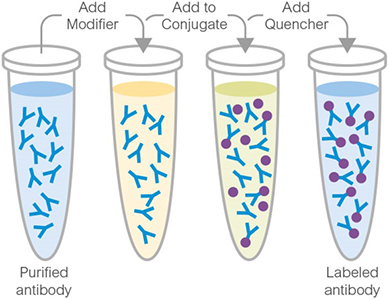
Abcam Lightning-Link® Conjugation Kit

主要特色

  • 操作極為快速,動手操作時間僅約 30 秒,全程約 20 分鐘即可完成。
  • 全程無需純化步驟,抗體回收率可達 100%,標定完成後即可直接應用於 Western Blot、ELISA、IHC 等實驗,無需額外處理。
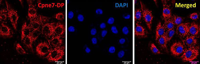
Immunocytochemistry. Lee, Yoon Seon, et al used Cy5® Conjugation Kit (Fast) - Lightning-Link® (ab188288) as part of examining the translocation and localization of newly developed peptide derived from CPNE7 (Cpne7-DP) . They used the kit to conjugate Cy5® to Cpne7-DP oligopeptide for use in immunocytochemistry. The intracellular distribution of Cpne7-DP is shown after odontoblastic MDPC-23 cells were treated with Cy5-labeled Cpne7-DP (10 μg/mL).
Immunocytochemistry. Lee, Yoon Seon, et al used Cy5® Conjugation Kit (Fast) - Lightning-Link® (ab188288) as part of examining the translocation and localization of newly developed peptide derived from CPNE7 (Cpne7-DP) . They used the kit to conjugate Cy5® to Cpne7-DP oligopeptide for use in immunocytochemistry. The intracellular distribution of Cpne7-DP is shown after odontoblastic MDPC-23 cells were treated with Cy5-labeled Cpne7-DP (10 μg/mL). Image from Lee et al., Materials, 13(20):4618; doi: 10.3390/ma13204618. Reproduced under the Creative Commons license https://creativecommons.org/licenses/by/4.0/
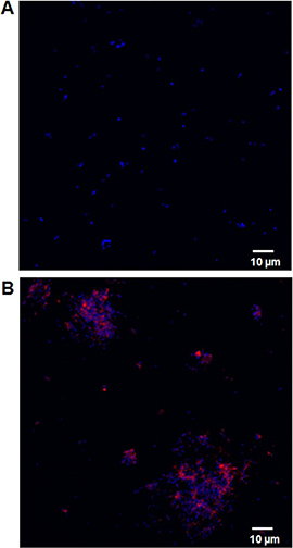
Immunocytochemistry/ Immunofluorescence. Dassanayake, Rohana P et al used ab188288 as part of examining the antimicrobial activity of bovine NK-lysin-derived peptides. They used the kit to conjugate Cy5® to NK2A peptide for use in confocal microscopy. H. somni was incubated with unconjugated Cy5 (A) or 20 μM NK2A-Cy5 conjugate for 30 min and bacterial nuclear contents were stained with DAPI. NK2A-Cy5 conjugate is shown in red and DAPI is shown in blue.
Immunocytochemistry/ Immunofluorescence. Dassanayake, Rohana P et al used ab188288 as part of examining the antimicrobial activity of bovine NK-lysin-derived peptides. They used the kit to conjugate Cy5® to NK2A peptide for use in confocal microscopy. H. somni was incubated with unconjugated Cy5 (A) or 20 μM NK2A-Cy5 conjugate for 30 min and bacterial nuclear contents were stained with DAPI. NK2A-Cy5 conjugate is shown in red and DAPI is shown in blue. Image from Dassanayake RP et al., PloS One, 12(8):e0183610. Fig 4.; doi:10.1371/journal.pone.0183610. Reproduced under the Creative Commons license https://creativecommons.org/licenses/by/4.0/
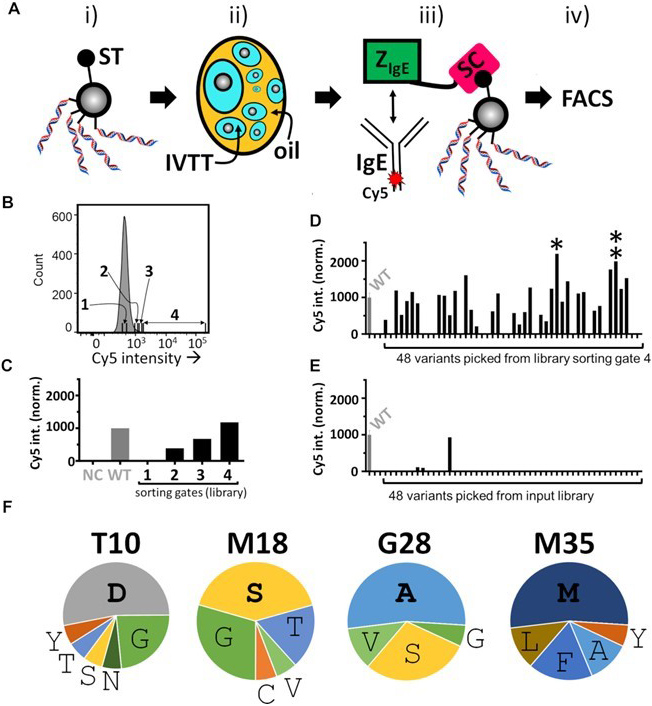
Flow Cytometry. Lindenburg, Laurens, et al used Cy5® Conjugation Kit (Fast) - Lightning-Link® (ab188288) as part of examining microemulsion-based bead display screening of the ZIgE SpliMLiB library. They used the kit to conjugate Cy5® to Native human IgE protein (Azide free) (ab65866) for use in FACS. (A) Schematic overview of a round of SpliMLiB-enabled directed evolution of ZIgE. SpliMLiB beads (i) were singly encapsulated in emulsion IVTT at 37°C for 1 h (ii), sufficient time to allow for both ZIgE-SpyCatcher variants' expression as well as for their SpyTag-SpyCatcher-mediated immobilisation on the bead surface, after which the emulsion was broken, and the washed beads were exposed to Cy5-labelled IgE (iii), followed by flow cytometric sorting of beads based on Cy5 signal (iv). (B) Representative histogram recorded during the flow cytometric sorting of SpliMLiB ZIgE library beads. The range of fluorescence intensity used for each of the sorting gates 1-4 is indicated. (C) Analysis of pooled, recovered and subcloned DNA from the sorting gates set out in panel B. DNA was used to express protein in IVTT under bulk, i.e. non-emulsion conditions, in the presence of SpyTag-functionalised microbeads. The microbeads, having captured the SpyCatcher fusion proteins, were then incubated with 200 nM IgE-Cy5 and analysed by flow cytometry. Cy5 fluorescence intensity was normalised to a sample prepared from beads exposed to purified ZIgEwild-type-SpyCatcher protein (WT, grey bar). Negative control (NC) was beads not exposed to any ZIgE-SpyCatcher protein. (D) Analysis of bacterially expressed & purified variants derived from the stringently sorted library output from FACS sorting gate 4. Beads that had been bound with ZIgE-SpyCatcher variants were incubated with 200 nM IgE-Cy5 and analysed by flow cytometry. ZIgEwild-type-SpyCatcher (labelled WT) was included as control and was used to normalise all fluorescent values. The variant showing the highest Cy5 median signal (variant 33, marked by a single asterisk) and second highest (variant 44, marked by a double asterisk) signal were taken forward for further analysis. (E) As panel D, except for 48 randomly picked clones derived from the unsorted SpliMLiB input library beads. (F) Frequencies of amino acids encountered in selected variants displaying a higher binding signal than ZIgEwild-type-SpyCatcher (17 in total). The most frequent amino acid at each position is indicated in bold to emphasise it.
Flow Cytometry. Lindenburg, Laurens, et al used Cy5® Conjugation Kit (Fast) - Lightning-Link® (ab188288) as part of examining microemulsion-based bead display screening of the ZIgE SpliMLiB library. They used the kit to conjugate Cy5® to Native human IgE protein (Azide free) (ab65866) for use in FACS. (A) Schematic overview of a round of SpliMLiB-enabled directed evolution of ZIgE. SpliMLiB beads (i) were singly encapsulated in emulsion IVTT at 37°C for 1 h (ii), sufficient time to allow for both ZIgE-SpyCatcher variants' expression as well as for their SpyTag-SpyCatcher-mediated immobilisation on the bead surface, after which the emulsion was broken, and the washed beads were exposed to Cy5-labelled IgE (iii), followed by flow cytometric sorting of beads based on Cy5 signal (iv). (B) Representative histogram recorded during the flow cytometric sorting of SpliMLiB ZIgE library beads. The range of fluorescence intensity used for each of the sorting gates 1-4 is indicated. (C) Analysis of pooled, recovered and subcloned DNA from the sorting gates set out in panel B. DNA was used to express protein in IVTT under bulk, i.e. non-emulsion conditions, in the presence of SpyTag-functionalised microbeads. The microbeads, having captured the SpyCatcher fusion proteins, were then incubated with 200 nM IgE-Cy5 and analysed by flow cytometry. Cy5 fluorescence intensity was normalised to a sample prepared from beads exposed to purified ZIgEwild-type-SpyCatcher protein (WT, grey bar). Negative control (NC) was beads not exposed to any ZIgE-SpyCatcher protein. (D) Analysis of bacterially expressed & purified variants derived from the stringently sorted library output from FACS sorting gate 4. Beads that had been bound with ZIgE-SpyCatcher variants were incubated with 200 nM IgE-Cy5 and analysed by flow cytometry. ZIgEwild-type-SpyCatcher (labelled WT) was included as control and was used to normalise all fluorescent values. The variant showing the highest Cy5 median signal (variant 33, marked by a single asterisk) and second highest (variant 44, marked by a double asterisk) signal were taken forward for further analysis. (E) As panel D, except for 48 randomly picked clones derived from the unsorted SpliMLiB input library beads. (F) Frequencies of amino acids encountered in selected variants displaying a higher binding signal than ZIgEwild-type-SpyCatcher (17 in total). The most frequent amino acid at each position is indicated in bold to emphasise it. Image from Lindenburg, Laurens, et al., Nucleic acids res., 8(11): e63; doi: 10.1093/nar/gkaa270. Reproduced under the Creative Commons license https://creativecommons.org/licenses/by/4.0/
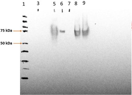
Western Blot. Mody, Karishma T., et al used Cy5® Conjugation Kit (Fast) - Lightning-Link® (ab188288) as part of examining the integrity of Bm86 protein postlabeling. They used the kit to conjugate Cy5® to R. micr-protein ladder, lane 5-Cy5-Bm86 protein, lane 7-Cy5-Bm86/ Rho-SV-140-C18 Supernatant, lane 8-Cy5-Bm86/Rho-SV-140-C18 pellet (first antibody 869 polyclonal sheep 1/5,000; second antibody monoclonal anti-goat/sheep 1/10,000). The Cy5-labeled Bm86 retained its native antigenicity as it was recognized by the antibodies in serum from a sheep immunized with the unlabeled antigen from a previous study. Image from Mody et al., Front bioeng biotechnol., 8:606652; doi: 10.3389/fbioe.2020.606652. Reproduced under the Creative Commons license https://creativecommons.org/licenses/by/4.0/
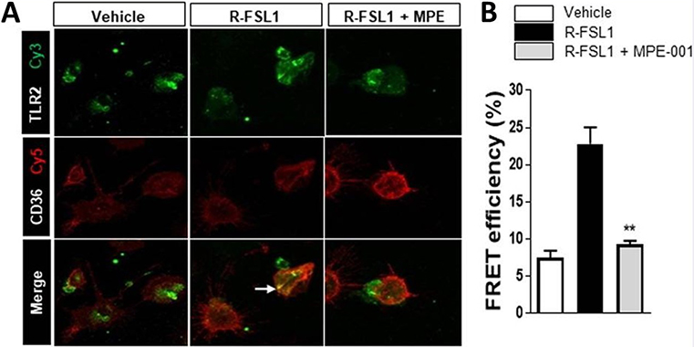
Fluorescence Resonance Energy Transfer (FRET). Mellal, Katia, et al used Cy5® Conjugation Kit (Fast) - Lightning-Link® (ab188288) as part of examining effects of MPE-001 on the CD36/TLR2 interaction. They used the kit to conjugate Cy5® to anti-CD36 antibody (ab80080) for use in FRET. Peritoneal MPs were stimulated with 300 ng/ml R-FSL1 in the presence of 10-7 M MPE-001 or vehicle. (A) MPE-001 disrupted the interaction between CD36 labeled with Cy5 (red) using the Cy5® Conjugation Kit (Fast) - Lightning-Link® (ab188288) and TLR2 labeled with Cy3 (green) using the Cy3® Conjugation Kit (Fast) - Lightning-Link® (ab188287) as assessed by FRET after 5 min stimulation with R-FSL1. (B) Percentage of energy transfer measured using LSM-700 confocal microscope (Zeiss). One-way ANOVA test with Newman-Keuls post-test for multiple comparison was performed. *P<0.05, **P<0.01 and ***P <0.001 vs R-FSL1. Data are shown as mean ±S.E.M.
Fluorescence Resonance Energy Transfer (FRET). Mellal, Katia, et al used Cy5® Conjugation Kit (Fast) - Lightning-Link® (ab188288) as part of examining effects of MPE-001 on the CD36/TLR2 interaction. They used the kit to conjugate Cy5® to anti-CD36 antibody (ab80080) for use in FRET. Peritoneal MPs were stimulated with 300 ng/ml R-FSL1 in the presence of 10-7 M MPE-001 or vehicle. (A) MPE-001 disrupted the interaction between CD36 labeled with Cy5 (red) using the Cy5® Conjugation Kit (Fast) - Lightning-Link® (ab188288) and TLR2 labeled with Cy3 (green) using the Cy3® Conjugation Kit (Fast) - Lightning-Link® (ab188287) as assessed by FRET after 5 min stimulation with R-FSL1. (B) Percentage of energy transfer measured using LSM-700 confocal microscope (Zeiss). One-way ANOVA test with Newman-Keuls post-test for multiple comparison was performed. *P<0.05, **P<0.01 and ***P <0.001 vs R-FSL1. Data are shown as mean ±S.E.M. Image from Mellal et al., Sci rep., 9(1):12903. doi: 10.1038/s41598-019-49472-8. Reproduced under the Creative Commons license https://creativecommons.org/licenses/by/4.0/

實驗注意事項

  • 樣本緩衝液 pH 值須介於 6.5–8.5,相容成分包括:Tris(濃度應 ≤50 mM / 0.6%)、BSA(≤0.1%)、甘油 (glycerol,≤50%)、疊氮化鈉 (Sodium Azide,≤0.1%)、磷酸鉀、氯化鈉、蔗糖、檸檬酸鈉、海藻糖等。緩衝液不可含有 Thimerosal (Thiomersal)、Proclin、Glycine、Arginine、Glutathione 等成分,以免干擾標定反應。更多樣本緩衝液細部條件說明,可參照下方表格或產品說明書。
  • 僅適用於已純化的抗體樣本;若樣本中含有其他蛋白質、胜肽或游離胺基分子,可能會競爭消耗標定試劑,影響標定效率。抗體樣本理想濃度為 1 mg/mL,若濃度不足(< 0.5 mg/ml)或過高(> 2 mg/ml),建議先進行濃縮或稀釋處理。
  • 標定完成的產物於 4°C 保存可穩定達 18 個月;若需長期保存,建議加入 50% 甘油作為冷凍保護劑,並存放於 -20°C。實際最適合的保存條件仍視個別抗體的穩定性而定,建議透過實驗確認。
Compatible buffer constituents
50mM / 0.6% Tris10.1% BSA250% glycerol
0.1% sodium azidePBSPotassium phosphate
Sodium chlorideHEPESSucrose
Sodium citrateEDTATrehalose
1 Tris buffered saline is almost always ≤ 50 mM / 0.6%
2 BSA can also interfere with the use of the conjugated antibody in tissue staining.
Incompatible buffer constituents
ThiomerosalProclinGlycine
ArginineGlutathioneDTT

套組內容物

Item3 x 10 µg100 µg3 x 100 µg1 mgStorage temperature
Cy5® mix3 vials1 vial3 vials1 vial-20°C
Modifier reagent1 vial1 vial1 vial1 vial-20°C
Quencher reagent1 vial1 vial1 vial1 vial-20°C

產品說明書

ab188288 Cy5® Conjugation Kit (Fast) - Lightning-Link®
返回頂端