Lightning-Link® FITC 抗體標定試劑盒

FITC Conjugation Kit - Lightning-Link®

貨號

ab102884

包裝

3 x 10 μg
100 μg
3 x 100 μg
1 mg

廠牌

Abcam

產品簡介

Abcam FITC Conjugation Kit - Lightning-Link® 提供了一種簡便快速的方法,可將 FITC 螢光染料 (fluorescein isothiocyanate, FITC) 標定至抗體或其他含有游離胺基的蛋白質、胜肽分子上,標定產物可直接應用於 Western Blot、流式細胞分析 (flow cytometry)、ELISA、免疫組織化學染色 (IHC) 等實驗。套組採用 Lightning-Link® 直接標定技術,僅需將修飾劑 (Modifier) 加入待標定抗體,再以此溶液回溶凍乾的 FITC 試劑小瓶,即可啟動標定反應;動手操作時間約 30 秒,3.5 小時左右即可完成標定,全程無需任何純化步驟,抗體回收率可達 100%。
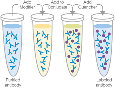
Abcam Lightning-Link® Conjugation Kit

主要特色

  • 操作簡便快速,套組採用 Lightning-Link® 直接標定技術,僅需將添加有 Modifier 的待標定抗體與凍乾的 FITC 試劑混合,即可透過離胺酸 (lysine) 等游離胺基進行共價標定,動手操作時間僅約 30 秒,整體反應時間在 3.5 小時左右(標定反應約 3 小時+中止反應 30 分鐘)。
  • 全程無需純化步驟,抗體回收率可達 100%,標定完成後即可直接應用於 Western Blot、Flow cytometry、ELISA、IHC 等實驗,無需額外處理。
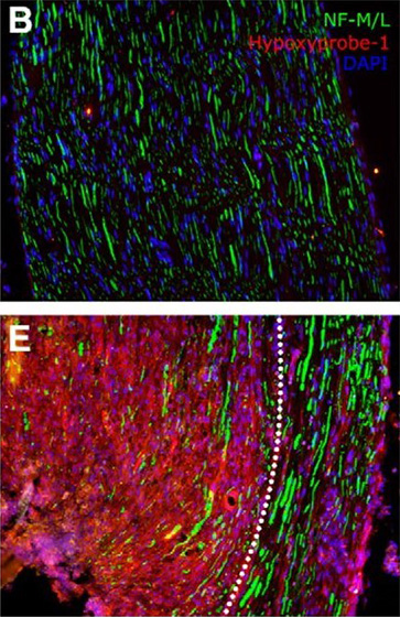
Immunocytochemistry. Lim, Tony KY, et al used FITC Conjugation Kit - Lightning-Link® (ab102884) as part of examining traumatic nerve injury. They used the kit to conjugate FITC to mouse anti-neurofilament 160/200 for use in immunohistochemistry.B, Contralateral nerves were stained for hypoxyprobe-1, DAPI, and neurofilament 160/200 (NF-M/L, axons). E, Ipsilateral nerves were subjected to the same staining. The area in proximity to the injury site is shown. Hypoxia, as shown by hypoxyprobe-1 staining, is observed in a region close to the injury site, in which axons, macrophages, and Schwann cells are all found to be hypoxic. This hypoxic region, in which nerve fibers were ligated, is delineated by the white dotted line. Areas outside of the dotted regions containing nonligated nerve fibers still have more hypoxyprobe deposition than contralateral nerves.
Immunocytochemistry. Lim, Tony KY, et al used FITC Conjugation Kit - Lightning-Link® (ab102884) as part of examining traumatic nerve injury. They used the kit to conjugate FITC to mouse anti-neurofilament 160/200 for use in immunohistochemistry.B, Contralateral nerves were stained for hypoxyprobe-1, DAPI, and neurofilament 160/200 (NF-M/L, axons). E, Ipsilateral nerves were subjected to the same staining. The area in proximity to the injury site is shown. Hypoxia, as shown by hypoxyprobe-1 staining, is observed in a region close to the injury site, in which axons, macrophages, and Schwann cells are all found to be hypoxic. This hypoxic region, in which nerve fibers were ligated, is delineated by the white dotted line. Areas outside of the dotted regions containing nonligated nerve fibers still have more hypoxyprobe deposition than contralateral nerves. Image from Lim, Tony KY, et al., J Neurosci., 35(8):3346-59, doi: 10.1523/JNEUROSCI.4040-14.2015. Reproduced under the Creative Commons license https://creativecommons.org/licenses/by/4.0/
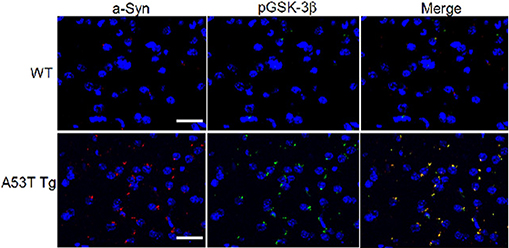
Immunohistochemistry (PFA perfusion fixed frozen sections). Wills, Jonathan, et al used FITC Conjugation Kit - Lightning-Link® (ab102884) as part of examining the aggregation and co-localization of α-Syn with pGSK-3β and p-Tau in the striatum of A53T α-Syn mutant mice (A53T Tg) and wild-type mice (WT). They used the kit to conjugate FITC to Anti-p-GSK-3β (pY216) antibody for use in immunohistochemistry (PFA perfusion fixed frozen sections). Right panels constitute merged image of left panels. Sections of striatum of A53T Tg and age-matched WT mice were stained with anti-α-Syn antibody conjugated to Texas Red (red) and anti-p-GSK3β conjugated to FITC (green). Nuclei were stained with DAPI (blue).
Immunohistochemistry (PFA perfusion fixed frozen sections). Wills, Jonathan, et al used FITC Conjugation Kit - Lightning-Link® (ab102884) as part of examining the aggregation and co-localization of α-Syn with pGSK-3β and p-Tau in the striatum of A53T α-Syn mutant mice (A53T Tg) and wild-type mice (WT). They used the kit to conjugate FITC to Anti-p-GSK-3β (pY216) antibody for use in immunohistochemistry (PFA perfusion fixed frozen sections). Right panels constitute merged image of left panels. Sections of striatum of A53T Tg and age-matched WT mice were stained with anti-α-Syn antibody conjugated to Texas Red (red) and anti-p-GSK3β conjugated to FITC (green). Nuclei were stained with DAPI (blue). Image from Wills, Jonathan, et al., PLoS One, 6(3): e17953; doi: 10.1371/journal.pone.0017953. Reproduced under the Creative Commons license https://creativecommons.org/licenses/by/4.0/
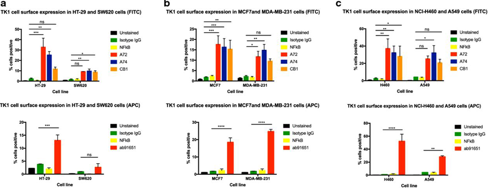
Flow Cytometry. Weagel, Evita G., et al used FITC Conjugation Kit - Lightning-Link® (ab102884) as part of examining thymidine kinase 1 in lung, breast, and colorectal malignancies. They used the kit to conjugate FITC to three custom and a commercial anti-human thymidine Kinase 1 (TK1) antibodies for use in flow cytometry. Membrane TK1 expression in of colon, breast, and lung cancer cell lines. Flow cytometry analysis of cell lines treated with anti-TK1 antibodies. a Quantification of TK1 expression on the cell membrane of HT-29 and SW620 cell lines stained with FITC or APC-conjugated anti-TK1 antibodies. b Quantification of TK1 expression on the cell membrane of MCF7 and MDA-MB-231 cell lines. The top bar graph shows MCF7 and MDA-MB-231 cell lines stained with FITC or APC-conjugated anti-TK1 antibodies. c Quantification of TK1 expression on the cell membrane of NCI-H460 and A549 cell lines stained with FITC or APC-conjugated anti-TK1 antibodies. Statistical analysis was performed by comparing the mouse isotype control fluorescent levels to those of A72, A74, CB1, or ab91651. *P ≤ 0.05; **P ≤ 0.005; ***P ≤ 0.001; ns = P > 0.05.
Flow Cytometry. Weagel, Evita G., et al used FITC Conjugation Kit - Lightning-Link® (ab102884) as part of examining thymidine kinase 1 in lung, breast, and colorectal malignancies. They used the kit to conjugate FITC to three custom and a commercial anti-human thymidine Kinase 1 (TK1) antibodies for use in flow cytometry. Membrane TK1 expression in of colon, breast, and lung cancer cell lines. Flow cytometry analysis of cell lines treated with anti-TK1 antibodies. a Quantification of TK1 expression on the cell membrane of HT-29 and SW620 cell lines stained with FITC or APC-conjugated anti-TK1 antibodies. b Quantification of TK1 expression on the cell membrane of MCF7 and MDA-MB-231 cell lines. The top bar graph shows MCF7 and MDA-MB-231 cell lines stained with FITC or APC-conjugated anti-TK1 antibodies. c Quantification of TK1 expression on the cell membrane of NCI-H460 and A549 cell lines stained with FITC or APC-conjugated anti-TK1 antibodies. Statistical analysis was performed by comparing the mouse isotype control fluorescent levels to those of A72, A74, CB1, or ab91651. *P ≤ 0.05; **P ≤ 0.005; ***P ≤ 0.001; ns = P > 0.05. Image from Weagel, Evita G., et al., Cancer cell international 18.1 (2018): 1-14. Reproduced under the Creative Commons license https://creativecommons.org/licenses/by/4.0/

實驗注意事項

  • 樣本緩衝液 pH 值須介於 6.5–8.5,相容成分包括:Tris(濃度應 ≤50 mM / 0.6%)、BSA(≤0.1%)、甘油 (glycerol,≤50%)、疊氮化鈉 (Sodium Azide,≤0.1%)、磷酸鉀、氯化鈉、蔗糖、檸檬酸鈉、海藻糖等。緩衝液不可含有 Thimerosal (Thiomersal)、Proclin、Glycine、Arginine、Glutathione 等成分,以免干擾標定反應。更多樣本緩衝液細部條件說明,可參照下方表格或產品說明書。
  • 僅適用於已純化的抗體樣本;若樣本中含有其他蛋白質、胜肽或游離胺基分子,可能會競爭消耗標定試劑,影響標定效率。抗體樣本理想濃度為 1 mg/mL,若濃度不足(< 0.5 mg/ml)或過高(> 2 mg/ml),建議先進行濃縮或稀釋處理。
  • 標定完成的產物於 4°C 保存可穩定達 18 個月;若需長期保存,建議加入 50% 甘油作為冷凍保護劑,並存放於 -20°C。實際最適合的保存條件仍視個別抗體的穩定性而定,建議透過實驗確認。
Compatible buffer constituents
50mM / 0.6% Tris10.1% BSA50% glycerol
0.1% sodium azidePBSPotassium phosphate
Sodium chlorideHEPESSucrose
Sodium citrateEDTATrehalose
1 Tris buffered saline is almost always ≤ 50 mM / 0.6%
Incompatible buffer constituents
ThiomerosalProclinGlycine
ArginineGlutathioneDTT

套組內容物

Item3 x 10 µg100 µg3 x 100 µg1 mgStorage temperature
FITC mix3 vials1 vial3 vials1 vial-20°C
Modifier reagent1 vial1 vial1 vial1 vial-20°C
Quencher reagent1 vial1 vial1 vial1 vial-20°C

產品說明書

ab102884 FITC Conjugation Kit - Lightning-Link®
返回頂端